What Is the Macula of the Eye and Why Is It Important for Central Vision?
Introduction
The macula (macula lutea) is a tiny area in the centre of the retina that determines our ability to see the world clearly and in detail. Thanks to the macula, we can read small print, drive a car, recognise faces, and distinguish the finest shades of colour.
When problems arise with the macula — whether age‑related changes, swelling, or a micro‑tear (macular hole) — the quality of central vision deteriorates significantly. This article explains where the macula is located, how it is structured, what functions it performs, and which diseases can affect it.
What Is the Macula and Where Is It Located?
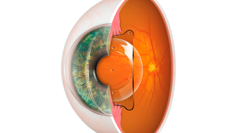
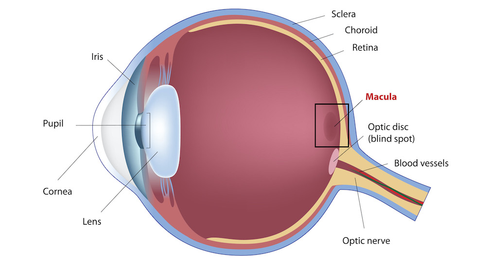
The macula is a specialised area of the retina, situated on the back wall of the eyeball (the fundus). It appears as a small yellowish area due to a high concentration of light‑sensitive pigment.
Key facts:
- Location: exact centre of the retina.
- Colour: yellowish spot (from Latin macula — “spot”).
- Function: converts light impulses into nerve signals that the brain turns into detailed images.
The Retina and the Ocular Fundus
The retina is the inner layer of the eye responsible for light perception. The macula occupies only a small part of it, but it provides the highest quality and sharpest vision. When an ophthalmologist examines the fundus, they evaluate the condition of the macula first.
Why Is the Macula So Important for Vision?
Without a healthy macula, everyday tasks become extremely difficult. This area of the retina gives us:
- High visual acuity — the ability to see fine details (letters, threads, road cracks).
- Colour perception — distinguishing shades and colour saturation.
- Central vision — the ability to focus directly on the object we are looking at.
If the macula is damaged, peripheral vision may remain good, but a person loses the ability to see what they are looking at directly.
Structure of the Macula
The macula is not just a “spot” — it is a complexly organised structure with several zones:
Fovea (Central Fovea)
Located in the very centre of the macula (diameter about 1.5 mm). This is the key area for sharpest vision. It has the highest density of cones — photoreceptors responsible for colour and detail.
Foveola
A depression inside the fovea (diameter about 0.35 mm). It appears as a small shiny reflex. The foveola provides the maximum possible visual acuity.
Foveal Avascular Zone (FAZ)
An area inside the fovea with no blood vessels. This allows light to reach the photoreceptors without any interference. On angiograms, this zone looks darker than the surrounding tissue.
Parafoveal Zone
The area surrounding the fovea (width about 0.5 mm). This is the thickest part of the retina.
Perifoveal Zone
The outer edge of the macula (diameter about 3.5 mm). A transitional area from central to peripheral vision.
Functions of the Macula
The macula performs several critically important tasks that go far beyond simple “seeing”.
High Resolution
Thanks to the high density of cones in the fovea, the macula allows us to distinguish the finest details: reading small fonts, seeing fabric textures, noticing tiny defects.
Colour Perception
Cones in the macula are specialised to detect different wavelengths of light (red, green, blue). Without a healthy macula, the world becomes dull and colours lose their saturation.
Motion Detection
The macula instantly transmits information about moving objects in the visual field to the brain. This is essential for driving, playing sports, and reacting to rapid changes in the environment.
Binocular Coordination (Stereopsis)
The macula is the fixation point for each eye. The brain combines signals from the right and left maculae into a single three‑dimensional image, allowing us to judge depth, distance, and volume.
Macular Diseases
Diseases of the macula severely reduce quality of life because they affect central vision directly. The most common conditions are listed below.
Age‑Related Macular Degeneration (AMD)
The most common macular disease, especially in people over 65. It involves progressive thinning or abnormal tissue growth in the centre of the retina. Peripheral vision usually remains, but reading, recognising faces, and watching television become impossible.
Macular Oedema
Fluid accumulation within the macula. The most common cause is diabetes mellitus (diabetic macular oedema). It can also occur with retinal vein occlusion, uveitis, after eye surgery, or as a side effect of certain medications. Symptoms: blurred vision, distortion of straight lines.
Macular Hole
A microscopic tear in the centre of the macula. Most often caused by traction (pulling) of the vitreous gel, which is normally attached to the retina. The disease has 4 stages:
- Stage 1 — may be asymptomatic.
- Stages 2–3 — distortion and vision loss appear.
- Stage 4 — significant loss of central vision (a dark spot in the centre).
Epiretinal Membrane (Macular Pucker)
A thin transparent membrane forms on the surface of the macula. Over time, it contracts and wrinkles the retina, causing image distortion (straight lines appear wavy). Early stages may be unnoticed, but as it progresses, reading becomes difficult and a dark spot may appear in central vision.
What to Do If You Suspect a Macular Problem
Any of the conditions described above requires immediate consultation with an ophthalmologist specialising in retinal diseases. Self‑diagnosis and delayed treatment can lead to irreversible central vision loss.
Symptoms to watch for:
- Reduced sharpness of central vision.
- Distortion of straight lines (windows, door frames, lines of text).
- A dark or hazy spot in the centre of your vision.
- Difficulty reading or recognising faces.
If you notice any of these symptoms — do not delay a visit to a specialist.
How Can Valenia Health Services Help?
Valenia Health Services specialises in arranging medical care for international patients in the best clinics of Barcelona. We work with leading ophthalmology centres in the city that successfully treat all types of macular pathology — from age‑related degeneration to macular holes and epiretinal membranes.
What we offer:
- Selection of the best retina specialist for your clinical case.
- Full organisation of diagnostics and treatment in Barcelona — no waiting lists, no language barriers.
- Professional medical translation and support at every stage.
- Transparent pricing — you pay only the clinic’s medical invoice, with no hidden fees.
We do not perform treatment ourselves, but we guarantee that you will see the best specialists in Spain, receive an accurate diagnosis, and get modern treatment.
Conclusion
The macula is a unique and extremely vulnerable structure of the eye. It makes our vision sharp, colourful, and detailed. Any macular disease, even at an early stage, requires specialist attention.
Timely diagnosis and proper treatment, organised in leading ophthalmology centres in Barcelona, can preserve vision for many years.